| Microglial activation, recruitment and phagocytosis as linked phenomena in ferric oxide nanoparticle exposure |
| From: PublishDate:2012-06-26 Hits: |
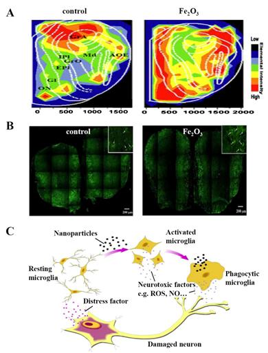
Microglia as the resident macrophage-like cells in the central nervous system (CNS) play a pivotal role in the innate immune responses of CNS. Understanding the reactions of microglia cells to nanoparticle exposure is important in the exploration of neurobiology of nanoparticles. Here we provide a systemic mapping of microglia and the corresponding pathological changes in olfactory-transport related brain areas of mice with Fe2O3-nanoparticle intranasal treatment. We showed that intranasal exposure of Fe2O3 nanoparticle could lead to pathological alteration in olfactory bulb, hippocampus and striatum, and caused microglial proliferation, activation and recruitment in these areas, especially in olfactory bulb. Further experiments with BV2 microglial cells showed the exposure to Fe2O3 nanoparticles could induce cells proliferation, phagocytosis and generation of ROS and NO, but did not cause significant release of inflammatory factors, including IL-1β, IL-6 and TNF-α. Our results indicate that microglial activation may act as an alarm and defense system in the processes of the exogenous nanoparticles invading and storage in brain. The micro-distribution mapping of iron in the rat brain after nanoparticle intranasal exposure was achieved by synchrotron radiation micro-beam X-ray fluorescence (SR-µXRF) at the Beijing Synchrotron Radiation Facility (BSRF) 4W1B beamline, with space resolution of several micrometer and detection limit of ng/g range. The incident beam was focused approximately 20 × 30 µm by a Pb collimator with two cross-slices. A monochromatic X-ray with photon energy of 16.5 keV was used to excite the samples. Samples were mounted on XYZ translation stages and the sample platform was moved by a 2D stepping motor along the X directions of 40 µm and Z directions of 60 µm for each step. In the study, we observed the activated microglial cells in olfactory bulb, hippocampus and striatum, accordingly with the storage route of Fe2O3 NP shown by SR-mXRF images and the alteration regions of the pathological reports. The most number of activated microglia were found in the olfactory nerve layer (ONL) and glomerular layer (GL), which was consistent with the transport pathway of Fe2O3 NPs along the olfactory nerve layers.
Biological effects of Fe2O3 nanoparticls on central nervous system. (A) Fe distribution in mouse olfactory bulb tested by SR-μXRF. (B) Immunofluorescence staining of CD11b in mouse olfactory bulb. Arrows in the box on the top right corner show CD11b-immunreactive microglia presenting “resting face” with a small cell body in the fine, ramified processes in control mice; in the Fe2O3 nanoparticls group mice, microglia present “activated face” with stouter cell processes. The Fe2O3 nanoparticls induced microglial proliferation and activation mainly concentrated in the olfactory nerve layer (ON) and glomerular layer (GL). (C) The mediation role of microglia for the protection or neurotoxicity of brain after intranasal exposure of Fe2O3 nanoparticls.
Article: Yun Wang§, Bing Wang§, Mo-Tao Zhu, Ming Li, Hua-Jian Wang, Meng Wang, Hong Ouyang, Zhi-Fang Chai, Wei-Yue Feng*, Yu-Liang Zhao, Microglial activation, recruitment and phagocytosis as linked phenomena in ferric oxide nanoparticle exposure. Toxicol. Lett. 2011, 205, 26-37. |
|
|
| Chinese
Science Highlights
Home /
Copyright © 2011 - 2012 Beijing Synchrotron Radiation Facility


